

A microscope is an instrument that allows people to see objects and details that are too small to be observed clearly with the naked eye. Microscopes have transformed science by revealing a hidden world of cells, microorganisms, tissues, crystals, and tiny structures. They are widely used in schools, laboratories, hospitals, research centers, and many other places where careful observation is important.
The most familiar type is the compound light microscope. It uses visible light and several lenses to enlarge an object placed on a glass slide. The lens closest to the specimen is called the objective lens, while the eyepiece is the part through which the viewer looks. Most microscopes have several objective lenses with different magnification levels. By rotating the nosepiece, users can move from a low-power view to a more detailed, high-power image.
Other important microscope parts include the stage, which supports the slide, and the focus knobs, which sharpen the image. The light source or mirror illuminates the specimen from below. A diaphragm controls how much light passes through the sample. Proper lighting and careful focusing are essential because an image may appear blurry, dark, or unclear when these parts are not adjusted correctly.
Microscopes can be used to examine plant cells, animal cells, bacteria, pond water, fibers, minerals, and many other materials. Scientists use them to study how living organisms grow, function, reproduce, and respond to disease. Medical professionals also use microscopes to analyze blood, tissue, and other samples that may help identify health problems.
More advanced instruments include electron microscopes, which use beams of electrons instead of visible light. They can produce extremely detailed images of viruses, cell structures, and materials at much higher magnifications. However, electron microscopes are larger, more expensive, and more complex than the light microscopes commonly found in classrooms.
Learning microscope vocabulary helps students understand how the instrument works and how scientific specimens are studied. A microscope word search can introduce useful terms such as lens, slide, stage, focus, specimen, magnification, objective, and eyepiece. Becoming familiar with these words makes laboratory instructions easier to follow and encourages students to explore the fascinating microscopic world with greater confidence and curiosity.
Related Word Searches:
Cell | Biology | Bacteria | DNA | Cell Division | Botany
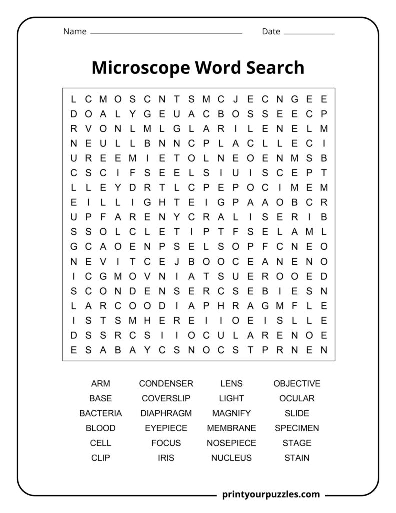
ARM, BASE, BACTERIA, BLOOD, CELL, CLIP, CONDENSER, COVERSLIP, DIAPHRAGM, EYEPIECE, FOCUS, IRIS, LENS, LIGHT, MAGNIFY, MEMBRANE, NOSEPIECE, NUCLEUS, OBJECTIVE, OCULAR, SLIDE, SPECIMEN, STAGE, STAIN
ARM – The curved or straight support connecting the microscope base to the head, providing structural stability. Users grip the arm when carrying the microscope to ensure safe transport.
BASE – The heavy, flat bottom of the microscope that provides stability and support. The base keeps the instrument steady during observation and often houses the light source.
BACTERIA – Microscopic single-celled organisms lacking a nucleus, visible only under microscopes. They appear as rods, spheres, or spirals and are essential subjects for microbiological study and research.
BLOOD – The red fluid circulating through bodies, containing cells visible under microscopes including red blood cells, white blood cells, and platelets, each with distinct shapes and functions.
CELL – The smallest unit of life, visible under microscopes. Cells contain genetic material and organelles, displaying various structures like membranes, nuclei, and cytoplasm when magnified for observation.
CLIP – Metal fasteners on the microscope stage that hold the slide securely in place during observation, preventing movement and ensuring the specimen remains properly positioned for viewing.
CONDENSER – A lens system beneath the microscope stage that concentrates and focuses light onto the specimen, improving image brightness, contrast, and overall clarity for better observation and detail.
COVERSLIP – A thin, flat piece of glass or plastic placed over a specimen on a slide to protect it, flatten it, and prevent evaporation during microscopic examination.
DIAPHRAGM – An adjustable opening below the microscope stage that controls the amount of light passing through the specimen, affecting image contrast, brightness, and depth of field for optimal viewing.
EYEPIECE – Also called the ocular lens, this is the lens at the top of the microscope that you look through, typically providing ten times magnification of the specimen.
FOCUS – The process of adjusting the distance between the objective lens and specimen to achieve a sharp, clear image by turning the coarse and fine adjustment knobs carefully.
IRIS – A type of diaphragm with overlapping metal plates that creates an adjustable circular opening, controlling light intensity and contrast by varying the aperture size beneath the stage.
LENS – A curved piece of glass or transparent material that bends light rays to magnify objects. Microscopes use multiple lenses working together to achieve high magnification levels.
LIGHT – Electromagnetic radiation in the visible spectrum used to illuminate specimens in optical microscopes, passing through or reflecting off samples to create magnified images for observation and study.
MAGNIFY – To enlarge the appearance of an object using lenses, making small details visible that cannot be seen with the naked eye, which is the primary function of microscopes.
MEMBRANE – A thin barrier surrounding cells and organelles, visible under microscopes with proper staining. Membranes control what enters and exits, appearing as delicate boundaries separating cellular compartments.
NOSEPIECE – The rotating turret that holds multiple objective lenses, allowing users to switch between different magnification powers by simply rotating the nosepiece to select the desired objective lens.
NUCLEUS – The control center of cells visible under microscopes as a dark, round structure. It contains DNA and regulates all cellular activities, growth, and reproduction processes.
OBJECTIVE – The primary lens closest to the specimen that gathers light and creates the initial magnified image. Microscopes typically have multiple objectives with different magnification powers on a rotating nosepiece.
OCULAR – Another term for the eyepiece lens through which the observer views the specimen. The ocular lens further magnifies the image created by the objective lens below.
SLIDE – A flat, rectangular piece of glass or plastic on which specimens are placed for microscopic examination. Slides hold samples in position for proper viewing and analysis.
SPECIMEN – A sample of material, tissue, or organism prepared and placed on a slide for examination under a microscope. Specimens can be living or preserved for scientific study.
STAGE – The flat platform on a microscope where the slide containing the specimen is placed. It often has clips to hold the slide securely in position during observation.
STAIN – A colored dye applied to specimens to enhance contrast and make specific structures more visible under the microscope. Different stains highlight different cellular components for easier identification.
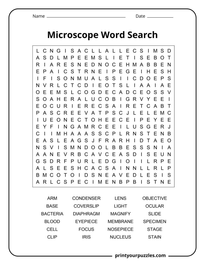
ARM, BASE, BACTERIA, BLOOD, CELL, CLIP, CONDENSER, COVERSLIP, DIAPHRAGM, EYEPIECE, FOCUS, IRIS, LENS, LIGHT, MAGNIFY, MEMBRANE, NOSEPIECE, NUCLEUS, OBJECTIVE, OCULAR, SLIDE, SPECIMEN, STAGE, STAIN
A microscope is a scientific instrument that uses lenses to magnify small objects or organisms, making them visible to the human eye. It’s essential for studying cells, bacteria, and tiny structures.
Light microscopes use visible light passing through lenses to magnify specimens. The objective lens creates an enlarged image, which the eyepiece lens further magnifies, producing the final visible image.
The main types include light microscopes for general use, electron microscopes for extremely high magnification, and stereo microscopes for three-dimensional viewing of larger specimens at lower magnification.
Light microscopes typically magnify between 40x and 1000x. Electron microscopes can achieve magnifications over 1,000,000x, allowing scientists to observe viruses, molecules, and atomic structures in remarkable detail.
Microscopes reveal cells, bacteria, blood components, tissue samples, microorganisms, plant structures, fungi, parasites, and countless other tiny specimens invisible to the naked eye, opening entire microscopic worlds.
Dutch spectacle makers Zacharias Janssen and his father Hans created the first compound microscope by placing multiple lenses in a tube, revolutionizing scientific discovery and our understanding of life.
In 1676, using his handcrafted single-lens microscope, Leeuwenhoek discovered “animalcules” (bacteria) in water samples, becoming the father of microbiology and opening an entirely new world of scientific exploration.
Unlike light microscopes limited to 2000x magnification, electron microscopes use electron beams instead of light, allowing scientists to visualize individual atoms, viruses, and molecular structures with extraordinary detail.
Simple clip-on lenses can transform smartphones into functional microscopes capable of 100x magnification, making scientific exploration accessible and affordable for students, hobbyists, and field researchers worldwide.
By visualizing microorganisms causing diseases, microscopes provided crucial evidence for germ theory in the 1800s, transforming medicine, hygiene practices, and our understanding of infection, ultimately saving millions of lives.